


.svg)
IsoMesh Scaffold
Guide cell alignment and securely position microtissues in a single, precision-manufactured scaffold.
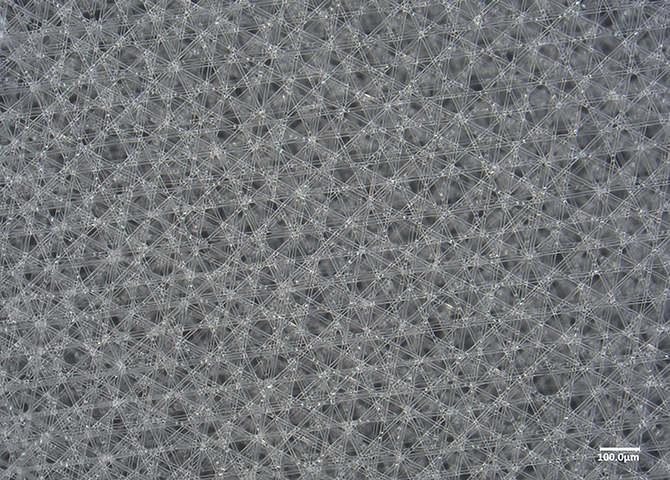
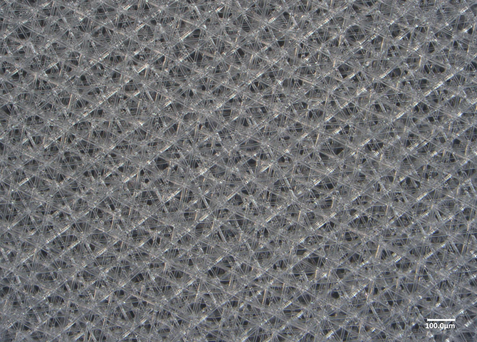
Microfibers are arranged in five evenly spaced orientations across 360°, resulting in a densely packed architecture. The isotropic design offers greater rigidity, reducing bending and deformation while improving cell seeding efficiency. This configuration has been successfully used to model subdermal skin in collaboration with L’Oréal.
Guide cell alignment and securely position microtissues in a single, precision-manufactured scaffold.
Microfibers are arranged in five evenly spaced orientations across 360°, resulting in a densely packed architecture. The isotropic design offers greater rigidity, reducing bending and deformation while improving cell seeding efficiency. This configuration has been successfully used to model subdermal skin in collaboration with L’Oréal.
KEY BENEFITS
Micron-Level Architectural Precision via Melt Electrowriting
Manufactured using melt electrowriting (MEW), the scaffolds deliver precise fiber diameter, fiber spacing, and alignment, with extreme precision for highly reproducible experiments.
Why it matters: Manufacturing consistency directly impacts experimental reproducibility, limiting comparability across studies and impact of results.
Provides a Backbone for Hydrogels
VivoTex MEW Scaffolds provide mechanical reinforcements to soft hydrogels that enable handling and manuverability
Why it matters: Cells prefer to grow in soft hydrogels. Handling, moving, and processing soft hydrogel constructs is challenging, and often introduces artifacts. MEW scaffolds provide a simple and powerful solution to workflows with soft hydrogel cultures.
Structural Cues for Cellular Patterning
Microfibers provide contact guidance cues that affect cell-cell interaction, cell elongation, migration, and organization along a defined axis, critical for modeling biological tissues. Alignment of microfibers provides directional guidance for cells and mechanical anisotropy that is common in many biological tissues such as muscles, tendons, bones, peripheral nerves, brain, and large blood vessels.
Why it matters: All biological tissue comprise fibrous microstructures that provide structural cues for cells and enable functional mechanical properties. Most hydrogel matrices fail to replicate the structural cues present in native ECM, limiting biological relevance.
Designed for Standard Lab Workflows
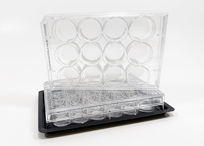
Scaffolds are designed to operate with standard well plates and other culture containers like petri dishes and chamber slides. They provide a solid and stable substrate for seeding, media exchange, staining, and imaging. No specialized equipment is required.
Why it matters: Enables advanced 3D culture without disrupting established lab protocols.
Applications & technical highlights
This Pattern Is Well-suited For:
The Mesh scaffold comprises 4,5, or 6 fiber directions equally distributed to create an an isotropic scaffold. These scaffolds are suited for membrane-like tissues such as skin and blood vessels
- Tissue engineering for novel approach methodologies (NAMs)
- Skin Equivalent models
Manufacturing Method:
Melt Electrowriting (MEW)
Architecture:
Aligned primary fibers
Format:
Compatible with standard well plate configurations
Use Case:
3D cell culture and microtissue integration
Mechanism of action
This scaffold functions through architecture-driven physical cues that regulate cell organization and microtissue positioning in 3D culture.
Aligned fibers provide anisotropic topographical guidance, directing cell adhesion, elongation, and migration along a defined axis.
Together, these features enable controlled cell alignment and reproducible microtissue placement while maintaining an open fiber network for media access and imaging. The scaffold’s deterministic fiber architecture, enabled by melt electrowriting, ensures consistent structural cues across experiments.
Configurable Formats
If this design does not meet your specific application, we can also collaborate to design custom scaffolds. This scaffold is available in multiple geometries and configurations to accommodate different experimental designs while maintaining consistent fiber alignment and architectural control.
In addition to flat formats compatible with standard well plates, the aligned fiber architecture can be fabricated in non-planar geometries, including tubular configurations. These formats are suited for models that require curvature, a defined lumen, or circumferential cell organization.
Experimental Relevance:
Scaffold geometry can influence cell organization, nutrient transport, and mechanical boundary conditions. Access to multiple form factors allows researchers to select a geometry that better matches their biological model without changing the underlying fiber architecture.
Use Cases May Include:
Flat scaffolds for aligned 3D cell culture and imaging in well-plate formats
- Tubular geometries for lumenized, circumferential, or axis-guided tissue models
- Alternative dimensions or layouts to match specific culture systems or devices
Configuration availability may vary. Contact Us at info@vivotex.com to discuss geometry options suitable for your experimental requirements or go here to learn more about our Custom Scaffold Solutions.
Material science overview
Manufactured via melt electrowriting, this scaffold features precisely placed, continuous microfibers with controlled diameter, spacing, and alignment. The resulting anisotropic architecture provides predictable pore geometry and mechanical behavior, while integrated catching fibers introduce localized structural features without reducing porosity. Deterministic fabrication ensures consistent material properties and reproducibility across experiments.
Research Studies Using This Pattern
technical documentation
How to Use This Product
For detailed guidance on handling, seeding, and culture workflows, visit the Getting Started page in our Support Center.