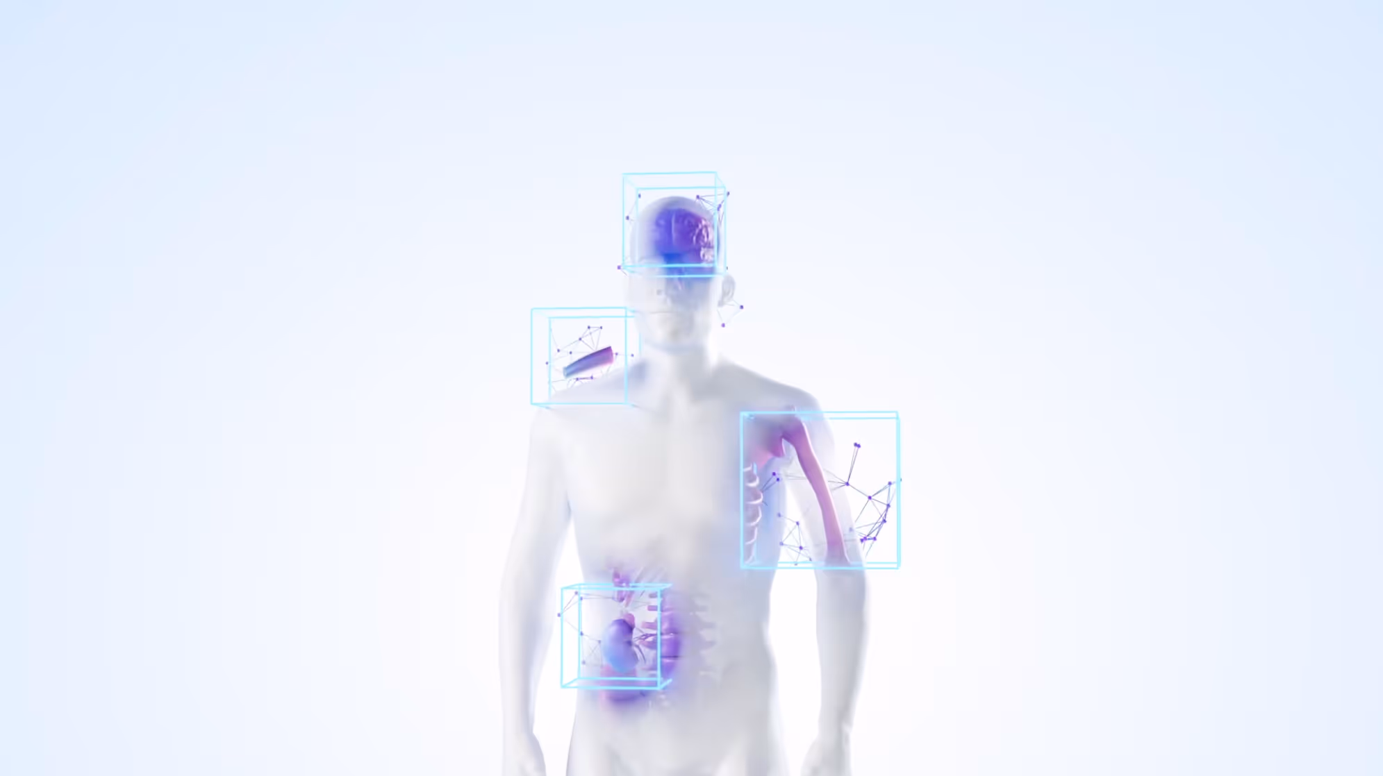
3d cell culture scaffolds

Backed by research, built for rigor, VivoTex 3D scaffolds enable trusted regenerative medicine studies, cancer research, and preclinical drug development.

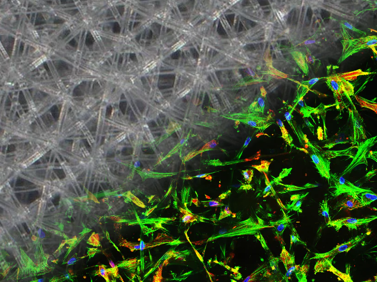
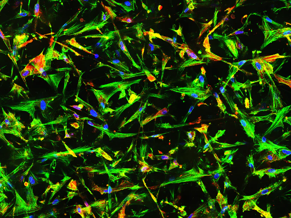
VivoTex 3D Scaffolds: Precision In Your Research

Designed with precision and adaptable to your research needs, VivoTex 3D scaffolds offer customizable architectures that replicate native tissue environments with consistency and control.

Need something specific? VivoTex scaffolds can be customized in architecture, porosity, and mechanics to match your experimental models. Learn more about customization.

Ready to try our scaffolds? Contact us and we'll send you a sample kit.

Technology  Highlights

Fully Customizable

Tunable architecture, fiber diameter, and porosity for diverse tissue models.

Precision at Scale

Lab-grade fabrication accuracy maintained in scalable production.

Sterile & Ready to Use

Pre-sterilized scaffolds integrate seamlessly into existing workflows.

Engineered Accuracy

Micron-level control over fiber placement for reproducible results.

OUR PRODUCT PORTFOLIO

Axis

Provides directional fiber guidance ideal for muscle, tendon, and neural models requiring cell alignment and organized growth.

View Product  >

Axis CF

Combines alignment cues with structural support, enabling stable, long-term studies of neural or musculoskeletal regeneration.

View Product  >

Lattice

Stabilizes 3D cultures for studying tissue organization, drug response, and regenerative models where structural support is essential.

View Product  >

Lattice CF

Secures organoids and spheroids for long-term culture, imaging, and high-resolution analysis without loss of structure.

View Product  >

IsoMesh

Replicates uniform, skin-like pore environments suited for wound healing, barrier tissue, and epithelial research.

View Product  >

How Researchers Use VivoTex Scaffolds

Scaffold Type

GEOMETRY/DESIGN

STRENGTHS

trade-offs

USE CASES/TISSUES

Rectilinear grid with uniform pores (e.g. ~150 µm)
Highly reproducible, easy seeding and imaging; strong baseline models.
Limited directional guidance for anisotropic tissues.
Epithelial barrier models, organoids, baseline comparisons
Same grid design with stabilizing “catching fibers" spanning pore gaps.
Maintains spheroid/organoid stability in long-term culture; reduces movement during handling.
Extra fibers can slightly obstruct imaging and migration paths.
Long-term tumor spheroid culture, drug testing in 3D aggregates.
Parallel fibers oriented in one direction; controlled fiber alignment.
Strong cues for cell alignment and elongation; mimics anisotropic ECM.
Less isotropic; biased growth may not suit mixed populations.
Muscle, tendon, neural, vascular alignment studies.
Aligned scaffold with added "catching fibers" cross-support fibers.
Combines directional guidance with scaffold stability; supports extended culture.
Higher fiber density may limit imaging clarity.
Long-term aligned tissue regeneration, mixed alignment + structure models.
Randomized, Multi-angle isotropic pattern fibers producing decagon / omnidirectional pore geometries.
Mimics heterogeneous ECM; uniform diffusion; versatile for non-directional growth.
Lacks alignment cues; less suited for tissues requiring order.
Skin/epithelial models, barrier testing, wound healing.

Box Pattern

GEOMETRY / DESIGN

Rectilinear grid with uniform pores (e.g. ~150 µm).

STRENGTHS

Highly reproducible, easy seeding and imaging; strong baseline models.

trade-offs

Limited directional guidance for anisotropic tissues.

USE CASES / TISSUES

Epithelial barrier models, organoids, baseline comparisons.

Box Pattern With Catching Fibers

GEOMETRY / DESIGN

Same grid design with stabilizing “catching” fibers spanning pore gaps.

STRENGTHS

Maintains spheroid / organoid stability in long-term culture; reduces movement during handling.

trade-offs

Extra fibers can slightly obstruct imaging and migration paths.

USE CASES / TISSUES

Long-term tumor spheroid culture, drug testing in 3D aggregates.

Aligned Pattern

GEOMETRY / DESIGN

Parallel fibers oriented in one direction; controlled fiber alignment.

STRENGTHS

Strong cues for cell alignment and elongation; mimics anisotropic ECM.

trade-offs

Less isotropic; biased growth may not suit mixed populations.

USE CASES / TISSUES

Muscle, tendon, neural, vascular alignment studies.

Aligned Pattern With Catching Fibers

GEOMETRY / DESIGN

Aligned scaffold with added cross-support fibers.

STRENGTHS

Combines directional guidance with scaffold stability; supports extended culture.

trade-offs

Higher fiber density may limit imaging clarity.

USE CASES / TISSUES

Long-term aligned tissue regeneration, mixed alignment + structure models.

Isotropic Pattern

GEOMETRY / DESIGN

Randomized, Multi-angle fibers producing decagon / omnidirectional pore geometries.

STRENGTHS

Mimics heterogeneous ECM; uniform diffusion; versatile for non-directional growth.

trade-offs

Lacks alignment cues; less suited for tissues requiring order.

USE CASES / TISSUES

Skin / epithelial models, barrier testing, wound healing.

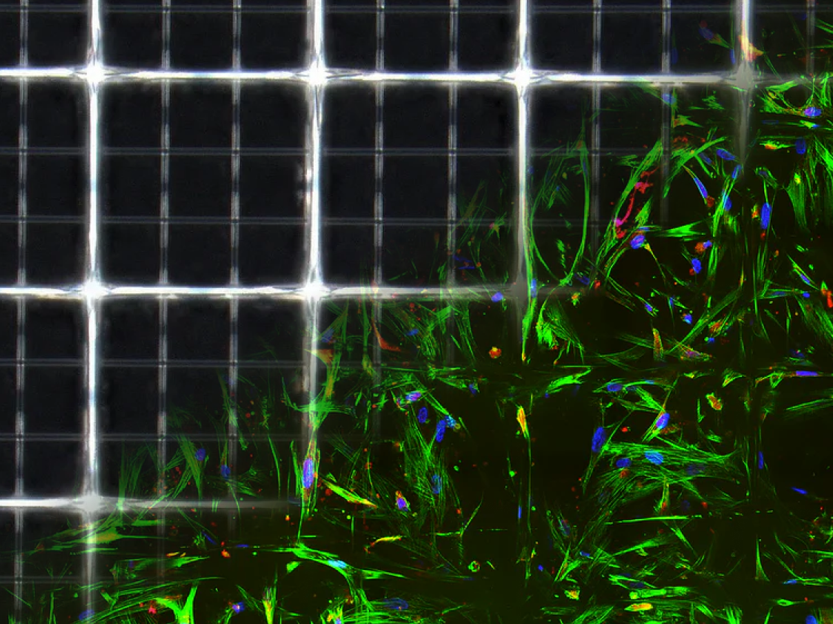
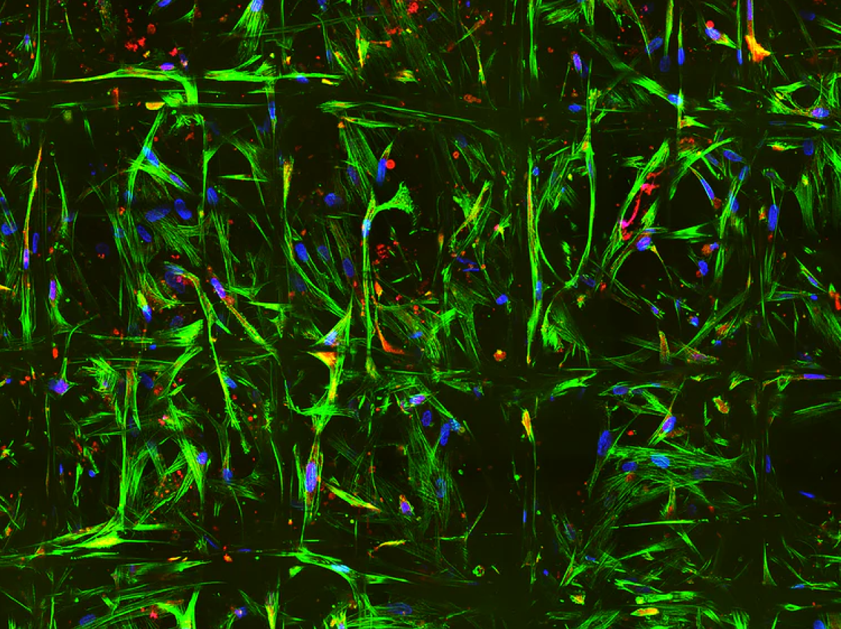
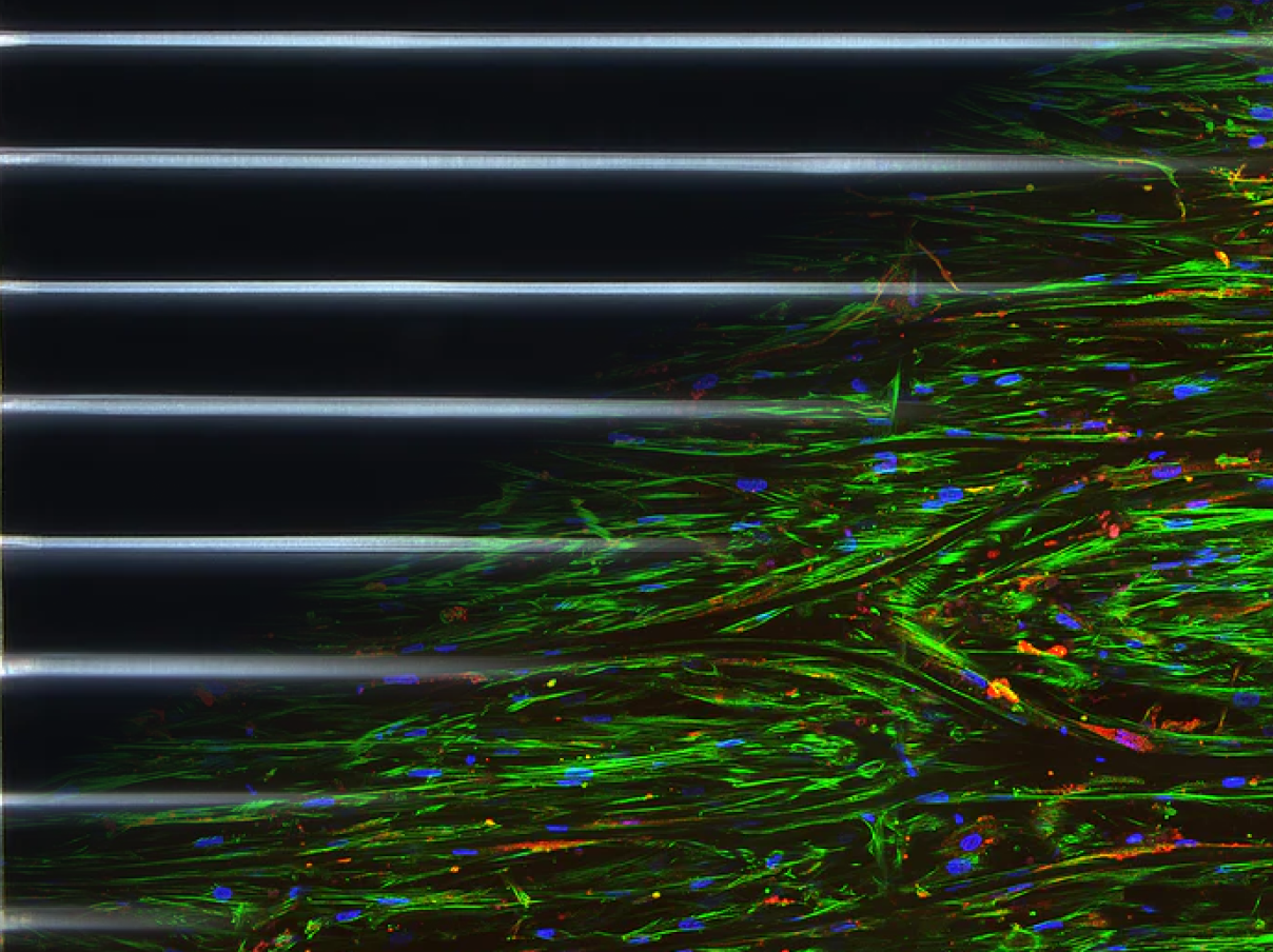
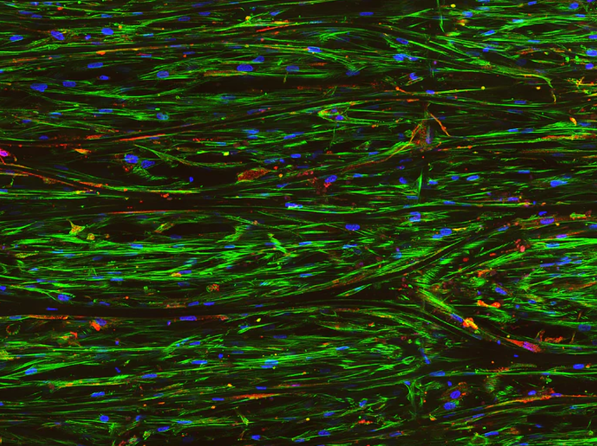
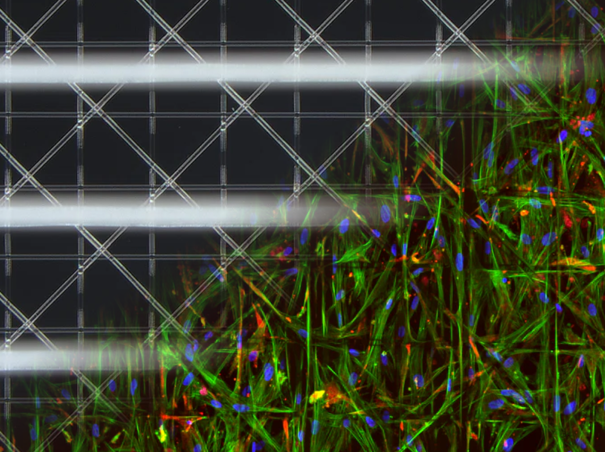
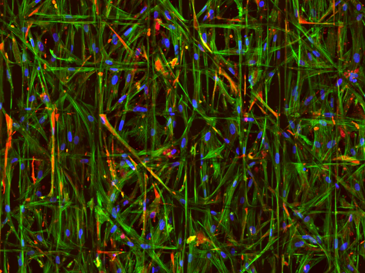
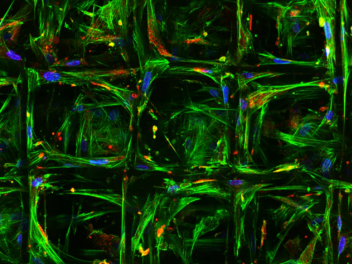
VivoTex Custom 3D Scaffolds: Tailored to Suit Your Needs

VivoTex scaffolds can be tailored to replicate specific tissue environments, giving researchers control over architecture, porosity, fiber diameter, and mechanical properties. This flexibility enables precise experimental models for diverse applications—from regenerative studies to oncology and drug discovery.

Ready to try our scaffolds? Contact us and we'll send you a sample kit.

PROBLEM

Current in-vitro models are costly, inconsistent, fail to replicate complex tissue environments, and are overly reliant on animal studies that lack human relevance, limiting reproducibility and predictive outcomes.

SOLUTION

VivoTex MEW scaffolds are reproducible, customizable, and biologically relevant, providing cost-effective human-relevant models that replicate complex tissues and deliver more predictive results.

Discover Our Advantage

IMPROVED OUTCOMES
Enables Nutrient Diffusion

Precisely spaced MEW fibers create open, interconnected structures that enable efficient nutrient & oxygen diffusion.

Supports Cell-to-cell Communication

Precisely controlled MEW micro-architecture facilitates cell–cell interactions that are critical for tissue organization & function.

Tunable Migration

Scaffold geometry, including fiber diameter, spacing, and orientation can be precisely tuned to guide cell migration & infiltration.

EASE OF USE
Ready to Use

MEW scaffolds are fabricated with defined geometry, delivered sterile & ready to use, with a team ready to support your success.

Reproducible Results

High-precision fabrication delivers uniform fiber placement & scaffold geometry, enabling consistent experimental outcomes.

Improved Handleability

Our scaffolds provide mechanical integrity & stability, improving handling during seeding, culture, & downstream analysis.

EXPLORE OUR TECHNOLOGY  >
Enables Nutrient Diffusion

Porous scaffolds enable efficient nutrient flow and waste removal for healthier, more active cells.

Supports Cell-to-Cell Communication

Precisely controlled MEW micro-architecture facilitates cell–cell interactions that are critical for tissue organization & function.

Tunable Migration

Scaffold geometry, including fiber diameter, spacing, and orientation can be precisely tuned to guide cell migration & infiltration.

Ready to Use

MEW scaffolds are fabricated with defined geometry, delivered sterile & ready to use, with a team ready to support your success.

Reproducible Results

High-precision fabrication delivers uniform fiber placement & scaffold geometry, enabling consistent experimental outcomes.

Improved Handleability

Our scaffolds provide mechanical integrity & stability, improving handling during seeding, culture, & downstream analysis.

The Competitive Landscape in 2D & 3D Cell Cultures

Hydrogels

Gel-based technologies provide ECM-like structure for cells to grow.

BioInks

Combination of cells and biopolymer gels “printed” into filaments.

Electrospun

3D printed membrane with random pattern at nano scale.

MEW

Polymeric scaffolds at micro scale provides higher precision and consistency.

Best Aligned
Cost to Customer
High Cost
High Cost
Low Cost
Neutral
Consistent Results
Weakness
Weakness
Neutral
Strength
Mimics Tissue Scale
Neutral
Neutral
Weakness
Strength
Mimics Human-Cellular  Architecture
Neutral
Weakness
Weakness
High Cost
Cell-to-cell Communication
Neutral
Neutral
Weakness
Strength
MEW

Polymeric scaffolds at micro scale provides higher precision and consistency.

Best Aligned
Cost to customer
Neutral
consistent results
Neutral
Mimics tissue scale
Neutral
mimics human-cellular architecture
Neutral
cell-to-cell communication
Neutral
Hydrogels

Gel-based technologies provide ECM-like structure for cells to grow.

Cost to customer
High Cost
consistent results
Weakness
Mimics tissue scale
Neutral
mimics human-cellular architecture
Neutral
cell-to-cell communication
Neutral
BioInks

Combination of cells and biopolymer gels “printed” into filaments.

Cost to customer
High Cost
consistent results
Weakness
Mimics tissue scale
Neutral
mimics human-cellular architecture
Weakness
cell-to-cell communication
Neutral
Electrospun

3D printed membrane with random pattern at nano scale.

Cost to customer
Low Cost
consistent results
Neutral
Mimics tissue scale
Weakness
mimics human-cellular architecture
Weakness
cell-to-cell communication
Neutral

Cell Morphology in 2D vs. 3D Culture

Comparison matrix across different 2d and 3d cell culture technologies/products
Advancing Hydrogel Performance

Hybrid MEW–hydrogel culture systems merge custom-engineered, cell-scale fibrous scaffolds with cell-compatible hydrogels, providing a highly porous structural backbone that enables easy handling of soft gels while better replicating the 3D cellular microenvironment and improving experimental reproducibility.

MEW in Peer-Reviewed Research

ADVANCED FUNCTIONAL MATERIALS

First Advanced Bilayer Scaffolds for Tailored Skin Tissue Engineering Produced via Electrospinning and Melt Electrowriting

Read Article >
SMALL

Biological Inspired Scaffolds for Heart Valve Tissue Engineering via Melt Electrowriting

Read Article >
JOURNAL OF THE MECHANICAL BEHAVIOR OF BIOMEDICAL MATERIALS

Melt Electrowritten Scaffold Architectures to Mimic Tissue Mechanics and Guide Neo-Tissue Orientation

Read Article >

Have Questions? Let's discuss Your Research Needs

Thank you! Your submission has been received!
Oops! Something went wrong while submitting the form.